Cat. No | MBA-015 |
Name | Rabbit anti-C-FOS Antibody |
中文名字 | 兔抗C-FOS抗体 |
Description | For induction of c-fos protein activity rats were injected with 1.0 ml of 1.5 M NaCl per 100 grams of body weight. Negative control rats were injected with the same volume of normal saline. The ImmunoStar c-fos antiserum was quality control tested using standard immunohistochemical methods. The antiserum demonstrates strongly positive labeling of rat paraventricular nucleus and supraoptic nucleus using indirect immunofluorescent and biotin/avidin-HRP techniques. No labeling was seen in negative control rats. Recommended primary dilutions for these methods are 1/4000-1/6000 in PBS/0.3% Triton X-100 – FITC Technique and 1/4000-1/6000 in PBS/0.3% Triton X-100 – biotin/avidin-HRP Technique. |
Clonality | Polyclonal |
Isotype | IgG |
Host | Rabbit |
Quantity/Volume | 50ul/100 µL |
State | Liquid Whole Serum/antibody |
Reacts With | Mouse, Rat |
Preabsorption Control | / |
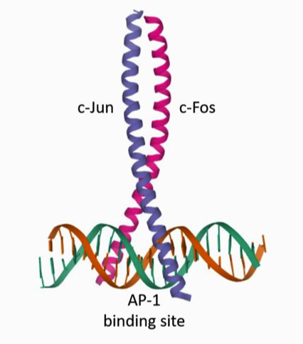
Alternate Names | Proto-oncogene c-Fos; Cellular oncogene; G0/G1 switch regulatory protein 7; Proto-oncogene protein; G0S7; p55; AP-1; FBJ murine osteosarcoma viral oncogene homolog, anti-CFOS |
RRID | AB_572267 |
Immunogen | This information is proprietary to Mabioway |
Gene Symbol | FOS |
Entrez Gene ID | Entrez Gene: 2353 Human |
NCBI Gene Aliases | / |
Sequence | >sp|P01100|FOS_HUMAN Protein c-Fos OS=Homo sapiens |
APPLICATION | |
Quality Control | For induction of c-Fos protein activity, rats were injected with 1.0 ml of 1.5 M NaCl per 100 grams of body weight. Negative control rats were injected with the same volume of normal saline. The ImmunoStar c-Fos antiserum was quality control tested using standard immunohistochemical methods. The antiserum demonstrates significant labeling of rat paraventricular and supraoptic nuclei using indirect immunofluorescent and biotin/avidin-HRP techniques. The antibody was also validated by challenge with the selective 5HT2a agonist TCB2 (10 mg/kg ip) with results showing massive numbers of cortical pyramidal cells of the TCB2 treated rats, consistent with the distribution of 5HT2a receptors. No labeling was seen in negative control rats. Specificity of the antiserum was demonstrated by blockage of staining in experimental rats by omission of c-Fos antibody or by substitution of antibody pre-incubated with synthetic peptide or the conjugate. |
Tissue | Rat brain hypothalamus (paraventricular and supraoptic nuclei) and cortex. |
Perfusion Fixation | • Fixative: 4% paraformaldehyde-0.05% glutaraldehye in 0.1M Phosphate buffer, pH 7.4; 500 mL over 20– 30 min. |
Absorption Control | / |
Sections | 50 µm vibratome |
Tissue Incubation | 18–24 hours at 2°–8°C |
Detection System | Use IF or Bn/Av-HRP reagents at dilutions recommended by the manufacturer. |
Suggested Dilution | 1/4,000–1/6,000 in PBS/0.3% Triton X-100 – Bn/Av-HRP immunohistochemistry |
NOTES | |
Special Instructions | It is recommended that the researcher perform a primary antibody dilution series using our dilution recommendations as a guideline. Note that a change in the fixation or buffering system from our protocol may change the configuration of the protein which could alter the reactivity with the tissue tested. |
Concentration | Not applicable. Antibody concentration is only relevant for purified antibodies. |
Storage | After reconstitution, use immediately or refrigerate at 2º–8ºC up to 2 days. For long term storage, aliquot and freeze at -15°C or lower. Avoid repeated freeze/thaw cycles. |


![Anti-NELL2 mabs [AT13E7.] (MA](http://www.mabioway.com/uploads/allimg/20240703/1-240F32152143S.png)
