Cat. No | MBA-0004 |
Name | Rabbit anti-5-HT (Serotonin) 2C Receptor Antibody |
中文名字 | 兔抗-5-HT 2C受体抗体,兔抗血清素抗体 |
Description | The ImmunoStar 5-HT2C receptor antiserum was quality control tested using standard immunohistochemical methods. The antiserum demonstrates strongly positive labeling of rat choroid plexus and hippocampus using indirect immunofluorescent and biotin/avidin-HRP techniques. Recommended primary dilutions are 1/500 – 1/1000 in PBS – Bn/Av-HRP technique. Intensification methods such as nickel will approximately double the dilution factor as recommended. |
Clonality | Polyclonal |
Isotype | IgG |
Host | Rabbit |
Quantity/Volume | 50ul/100 µL |
State | Liquid |
Reacts With | Mouse, Rat |
Preabsorption Control | / |
Alternate Names | 5-hydroxytryptamine receptor 2C; 5-HT2C; 5HT-1C; 5-HTR2C; 5-hydroxytryptamine (serotonin) receptor 2C, G protein-coupled, anti-5-HT 2C |
RRID | AB_572212 |
Immunogen | This information is proprietary to Mabioway |
Gene Symbol | Htr2c |
Entrez Gene ID | Entrez Gene: 15560 Mouse |
NCBI Gene Aliases | |
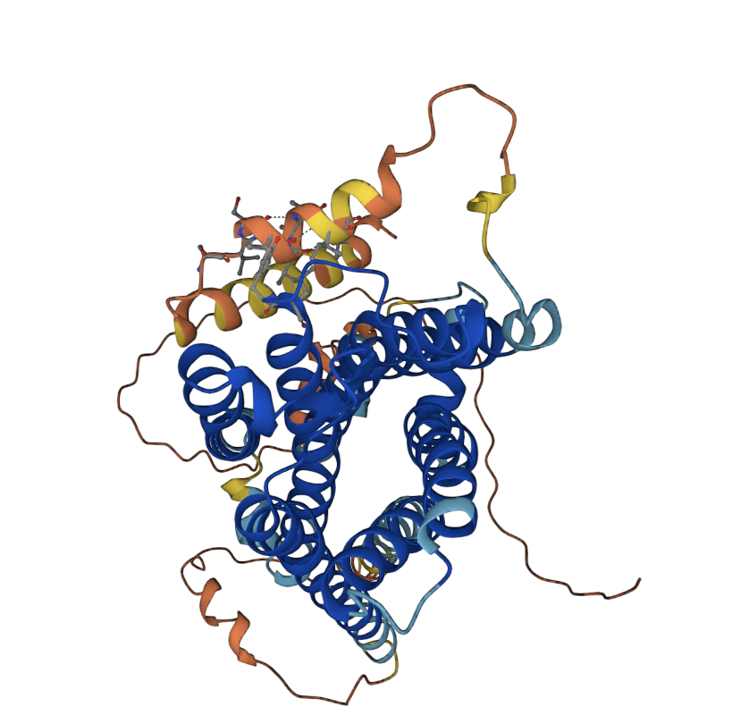
Sequence | >sp|P28335|5HT2C_HUMAN 5-hydroxytryptamine receptor 2C |
APPLICATION | |
Quality Control | The 5-HT2C receptor antiserum was quality control tested using standard immunohistochemical methods. The antiserum demonstrates significant labeling of rat choroid plexus and hippocampus using indirect immunofluorescent and biotin/avidin-HRP techniques. Intensification methods such as nickel will approximately double the dilution factor as recommended. Preincubation of the antibody with an excess of the synthetic peptide blocked staining. Immunohistochemical staining of rat brain correlates well with northern analysis, in situ hybridization and receptor autoradiography. The antibody was characterized by immunohistochemistry and western blot. Western blotting revealed a single band of approximately 70 kD. Due to the difficulty with receptor antibodies, western blot applications are not warranted and are included as specificity information only. |
Tissue | Rat cortex, hippocampus, hypothalamic nuclei |
Perfusion Fixation | • Fixative: 4% paraformaldehyde in 0.1M Phosphate buffer, pH 7.4; 500 mL over 20 min. |
Absorption Control | / |
Sections | 50 µm vibratome |
Tissue Incubation | 48 hours at 2°–8° C. |
Detection System | Use Bn/AV-HRP at dilutions recommended by the manufacturer |
Suggested Dilution | 1/300–1/600 in PBS - Bn/Av-HRP technique |
NOTES | |
Special Instructions | It is recommended that the researcher perform a primary antibody dilution series using our dilution recommendations as a guideline. Note that a change in the fixation or buffering system from our protocol may change the configuration of the protein which could alter the reactivity with the tissue tested. |
Concentration | Not applicable. Antibody concentration is only relevant for purified antibodies. |
Storage | Store at 2°–8°C until expiration date. |


![Anti-NELL2 mabs [AT13E7.] (MA](http://www.mabioway.com/uploads/allimg/20240703/1-240F32152143S.png)
