Cat.No | MBA-0002 |
Name | Rabbit anti-5-HT (Serotonin) 1A Receptor Antibody |
中文名字 | 兔抗-5-HT 1A受体抗体,兔抗血清素抗体 |
Description | The histochemical antibody for 5-HT1A receptor is generated in a Rabbit against synthetic peptide sequence corresponding to amino acids 294-312 of the rat 5-HT1A receptor. The antiserum is provided as 100 µl of affinity purified serum containing 1% BSA. Controls: The ImmunoStar 5-HT1A receptor antiserum was quality control tested using standard immunohistochemical methods. |
Clonality | Polyclonal |
Isotype | IgG |
Host | Rabbit |
Quantity/Volume | 50ul/100 µL |
State | Liquid |
Reacts With | Guinea Pig, Human, Monkey, Mouse, Rat |
Preabsorption Control | / |
Alternate Names | 5-hydroxytryptamine receptor 1A; ADRB2RL1; ADRBRL1; RAT5HT1A, 5-hydroxytryptamine (serotonin) receptor 1A, G protein-coupled, anti-5-HT 1A |
RRID | AB_572210 |
Immunogen | This information is proprietary to Mabioway |
Gene Symbol | Htr1a |
Entrez Gene ID | Entrez Gene: 100301563 Guinea Pig |
NCBI Gene Aliases | / |
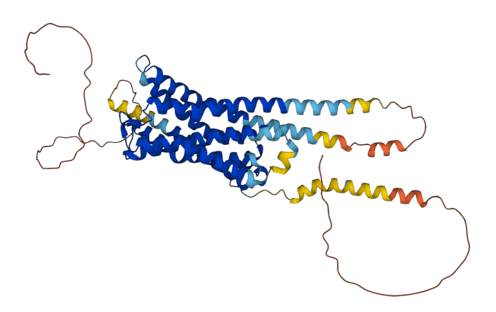
sequence | >sp|P08908|5HT1A_HUMAN 5-hydroxytryptamine receptor 1A |
APPLICATION | |
Quality Control | The 5-HT1A receptor antiserum was quality control tested using standard immunohistochemical methods. The antiserum demonstrates significant labeling of rat cortex, arcuate and hippocampus using indirect immunofluorescent and biotin/avidin-HRP techniques. Intensification methods such as nickel will approximately double the dilution factor as recommended. The antibody was characterized by immunohistochemistry and western blot. Western blot showed a single band of approximately 45 kD. Due to the difficulty with receptor antibodies, western blot applications are not warranted and are included as specificity information only. Preincubation of the antibody with an excess of the synthetic peptide blocked staining. Immunohistochemical staining of rat brain correlates well with Northern analysis, in situ hybridization and receptor autoradiography. |
Tissue | Rat cortex, arcuate and hippocampus |
Perfusion Fixation | • Fixative: 4% paraformaldehyde/0.05% glutaraldehyde in 0.1M Phosphate buffer, pH 7.4; 500 mL over 20 min. |
Absorption Control | / |
Sections | 50 µm vibratome. Cryosections are not recommended as it may disrupt the membrane. This antibody does not work with paraffin embedded tissues. |
Tissue Incubation | 48 hours at 2°–8° C. |
Detection System | Use Bn/AV-HRP at dilutions recommended by the manufacturer. For immunofluorescence stain wash in 1% sodium borohydride after the wash step following fixation in order to quench aldehyde sites and aid in suppressing background. See instructions online for western blot suggestions. |
Suggested Dilution | 1/200 –1/300 in PBS - Bn/Av-HRP detection. 1/100 or greater in Western Blot. Note: Use of Triton X-100 or other detergents is not recommended. If necessary, use only 0.03% T-X100 in the block only. No more detergent thereafter. Further penetration enhancement may be achieved by subsequent 5 min washes in 10, 25, 40, and 50% ethanol in PBS, followed by reversing wash order back down to PBS. |
NOTES | |
Special Instructions | It is recommended that the researcher perform a primary antibody dilution series using our dilution recommendations as a guideline. Note that a change in the fixation or buffering system from our protocol may change the configuration of the protein which could alter the reactivity with the tissue tested. |
Concentration | Not applicable. Antibody concentration is only relevant for purified antibodies. |
Storage | Store at 2°–8°C until expiration date. |


