Cat.No | MBA-0001 |
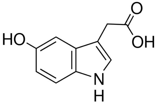
Name | Rabbit anti-5-HIAA (5-Hydroxyindoleacetic Acid) Antibody |
中文名字 | 兔抗5-HIAA抗体, 兔抗5-羟基吲哚乙酸抗体 |
Description | The antibody produces moderate labeling of raphe neurons in normal rat. In rats whose serotonergic system has been activated, staining intensity is increased to a maximum label. Recommended dilution of the antiserum is 1/4000-1/8000 for biotin-streptavidin/HRP technique. |
Clonality | Polyclonal |
Isotype | IgG |
Host | Rabbit |
Quantity/Volume | 50ul/100 µL |
State | Liquid Whole Serum/antibody |
Reacts With | Rat, Tritonia Diomedea (Sea Slug),Mollusca |
Preabsorption Control | / |
Alternate Names | anti-5-HIAA |
RRID | AB_572208 |
Immunogen | This information is proprietary to Mainway |
Gene Symbol | SLC6A4 |
Entrez Gene ID | 6532 |
NCBI Gene Aliases | 5-HTT,5-HTTLPR,5HTT, HTT,OCD1,SERT,hSER |
sequence | 54-16-0 |
APPLICATION | |
Quality Control | The antibody produces significant labeling of raphe neurons in normal rat. In rats whose serotonergic system has been activated, staining intensity is increased to a significant label. Recommended dilutions of the antiserum are 1/4,000–1/8,000 for indirect immunofluorescence and for biotin-streptavidin/HRP technique. The specificity of the antiserum was evaluated using a model system of gelatin-indole plugs by a method similar to published procedures (Schipper and Tilders, 1983). Results showed that the 5-HIAA antibody dose dependently stained 5-HIAA but did not stain any concentration of 5-HT or 5-HTP. The antiserum was also tested by preadsorption at 25 mg/mL with various BSA conjugates. While preadsorption with 5-HIAA conjugate completely eliminates immunolabeling, preadsorption with conjugates of 5-HT, 5-HTP and dopamine had no effect on staining intensity or distribution of stain. |
Tissue | Rat dorsal and median raphe neuronal cell bodies. Serotonergic system may be activated by salt loading which is achieved by 2% NaCl placed in drinking water for 48 hours prior to perfusion. |
Perfusion Fixation | • Fixation: 4% paraformaldehyde in 0.1M phosphate buffer, pH 7.4; 500 mL over 20 min. |
Absorption Control | |
Sections | 10um cryostat or 50um vibratome |
Tissue Incubation | 18–24 hours at 2°–8°C. |
Detection System | Use IF or Bn-AV/HRP reagents at dilutions recommended by the manufacturers |
Suggested Dilution | 1/4,000–1/8,000 in PBS/0.3% Triton X-100 – Bn-AV/HRP immunohistochemistry |
NOTES | |
Special Instructions | It is recommended that the researcher perform a primary antibody dilution series using our dilution recommendations as a guideline. Note that a change in the fixation or buffering system from our protocol may change the configuration of the protein which could alter the reactivity with the tissue tested. |
Concentration | Not applicable. Antibody concentration is only relevant for purified antibodies. |
Storage | Store at-20°C until expiration date. |


